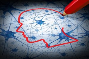
Psychophysiology is the scientific study of the connection between the mind (psychology) and the body (physiology). Psychophysiology is based on the premise that there is two-way communication between the mind and the body. Thus, physical changes in the body can affect psychological responses in the mind and psychological experiences may be reflected in physiological measures.
Psychophysiology has been used extensively in the field of sport and exercise psychology (SEP). In sport psychology (SP), psychophysiological measures have been used with athletes to understand physiological variables that are predictive of performance and to use this understanding to improve performance. In exercise psychology, scientists have used psychophysiological measures to provide physical evidence of the effects of exercise on a variety of psychological experiences including cognitive function and brain health, stress and anxiety, affect, arousal, and sleep.
Academic Writing, Editing, Proofreading, And Problem Solving Services
Get 10% OFF with 24START discount code
Psychophysiological Measures
The psychophysiological measures relevant to SEP are constantly evolving as technological advances allow for the use of noninvasive measures and theory advances our understanding of potential mechanisms of relevant outcomes. The measures described here are commonly used in SEP research.
Magnetic Resonance Imaging
 By utilizing finely tuned magnetic pulses that disrupt the orientation of atoms in the brain, magnetic resonance imaging (MRI) is used to assess cerebral structure. MRI is capable of making multiple assessments of the brain at varying depths and in different directional planes (sagittal, coronal, transverse). This makes it possible to assess the cerebral volume of specific areas of the brain. Exercise psychologists have used MRI to assess changes in cerebral volume in relation to physical activity (PA) in clinical (e.g., Alzheimer’s disease) and nonclinical populations.
Functional Magnetic Resonance Imaging
The brain requires a supply of oxygenated blood in order to support neuronal activation. In areas of the brain where activation has occurred, blood flow is increased to compensate for the consumption of oxygen and other vital nutrients. The increased blood flow results in a higher concentration of oxygenated hemoglobin (the protein that transports oxygen) in the specific areas of activation relative to less active areas. Functional magnetic resonance imaging (fMRI) is able to detect the different magnetic properties of oxygenated and deoxygenated hemoglobin in blood, and this blood-oxygen-level-dependent (BOLD) response is used to infer neural activation, which is interpreted as indicative of cognitive activity.
Heart Rate and Heart Rate Variability
Heart rate (HR) provides a simple measure of arousal and affect. An increase in HR has been interpreted as indicative of an increase in psychological arousal or a change in mood. Assessing HR can be done manually by palpating the wrist or carotid artery and counting the number of beats per minute (BPM), by using a heart monitor, or by measuring electrocardiographic activity using electrodes
Heart rate variability (HRV) refers to variations in the time between heartbeats (which is also referred to as the interbeat interval [IBI]). HRV has been used to make inferences about the control of HR by the autonomic nervous system (ANS). In particular, in the SEP literature, HRV is interpreted relative to high-frequency (parasympathetic activity) and low-frequency (both sympathetic and parasympathetic activity) components.
Electroencephalography
Electroencephalography (EEG) (also referred to as electroencephalographic activity) is the process of recording electrical activity across the scalp. The supposition is that the EEG activity is
indicative of the neural activity taking place in the brain. EEG is described as being high in temporal resolution because observed activity is related to current cognitive processes but low in spatial resolution because data represents the firing of a large number of neurons in a general area that may or may not be located near the recording location. EEG recordings may be used to assess either spontaneous activity or event-related potentials (ERPs). These constructs are described further in the entry titled Electroencephalograph (EEG).
Electromyography
Electromyography (EMG) is used to measure skeletal muscle activity (sometimes referred to as tension) by taking measures of electrical activity either at the skin using surface electrodes or directly from the muscle using intramuscular electrodes. EMG measures have been used to identify the timing of muscle activation and to provide an indication of levels of arousal, stress, and affect.
Galvanic Skin Response
Galvanic skin response (GSR) is measured by assessing skin conductance (SC). An electrical stimulus with an extremely small voltage is applied between two electrodes adhered to the skin, and the conductance between them is recorded. GSR is directly reflective of moisture level and is interpreted as being indicative of sympathetic nervous system (SNS) activity and arousal.
Cortisol
Cortisol is also referred to as the “stress hormone” and is released when the hypothalamicpituitary-adrenal (HPA) gland axis is stimulated. When a person is exposed to a stressful stimulus, the brain (i.e., hypothalamus and pituitary gland) signals the adrenal glands to release cortisol from the adrenal glands to prepare the body for fight or flight. When it senses the end of the stressor, the brain begins the reuptake processes of removing the cortisol from the blood stream, which returns the body to homeostasis. Cortisol levels are indicative of stress and can be assessed in peripheral blood and in saliva.
In addition to the psychophysiological measures described here, researchers in the field of SEP have also used measures of critical flicker fusion, acoustic startle eye blink responses, neurotransmitters (e.g., serotonin, dopamine), hormones (e.g., epinephrine), and body temperature to enhance our understanding of mind and body interactions.
References:
- Aubert, A. E., Seps, B., & Beckers, F. (2003). Heart rate Lewis, M. J., & Short, A. L. (2010). Exercise and cardiac regulation: What can electrocardiographic time series tell us? Scandinavian Journal of Medicine & Science in Sports, 20(6), 794–804. doi: 10.1111/j.1600-0838.2010.01150.x
- Weinstein, A. M., & Erickson, K. I. (2011). Healthy body equals healthy mind. Generations, 35(2), 92–98.
- Zaichkowsky, L. (Ed.). (2012). Psychophysiology and neuroscience [Special issue]. Journal of Clinical Sport Psychology, 6(1). variability in athletes. Sports Medicine, 33(12), 889–919.
- Gatti, R., & De Palo, E. F. (2011). An update: Salivary hormones and physical exercise. Scandinavian Journal of Medicine & Science in Sports, 21(2), 157–169. doi:10.1111/j.1600-0838.2010.01252.x
See also: